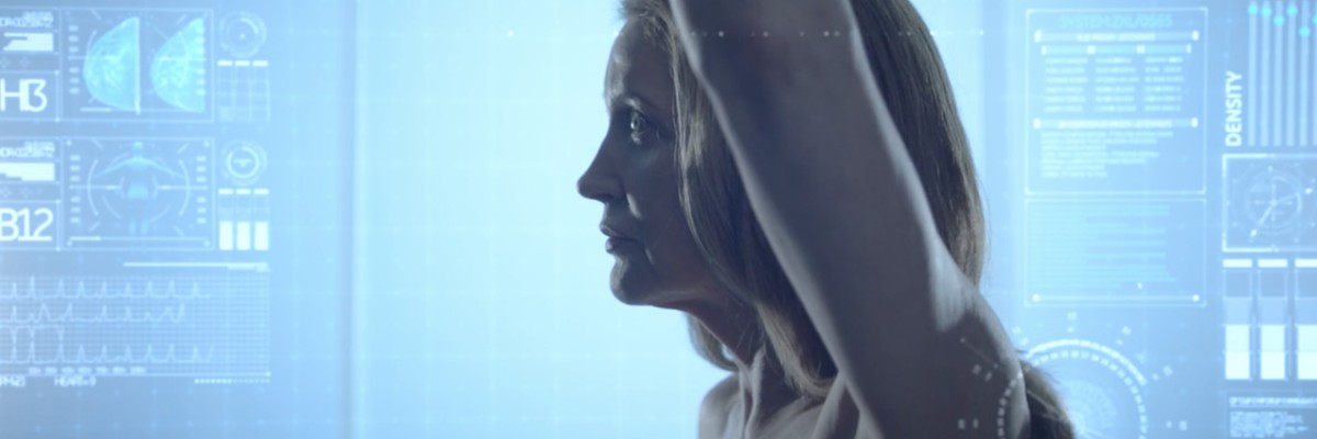


The healthcare community continues to strive for early detection of breast cancers — primarily through screening mammography, which is known to be an effective method for detecting early breast cancers and has been shown to reduce breast cancer-specific mortality[1]. Other powerful imaging modalities such as breast ultrasound and magnetic resonance imaging (MRI) are also frequently utilized for some cases in breast cancer detection. To aid clinicians in finding these cancers, artificial intelligence (AI)-based tools have been introduced and show great promise in increasing diagnostic accuracy, as well as creating substantial improvements in productivity[2].
Interest in AI in healthcare is on the rise, with $4 billion invested into the sector in 2019, up from $2.7 billion in 2018[3]. While many AI-based solutions have already been adopted into healthcare environments to support specific processes and tasks, new AI solutions and machine learning algorithms have been introduced in breast imaging to assist radiologists in the detection of breast cancers. Deep learning, which is a subset of AI, utilizes neural network models with many levels of features or variables that help predict patient outcomes. Using these deep-learning tools and also radiomics, which is the detection of clinically relevant features in imaging data beyond what can be perceived by the human eye,[4] clinicians are aided in oncology-oriented image analysis.
In the latest webinar of the Beyond Breast Imaging series hosted by GE Healthcare, a panel of global experts in breast imaging came together to discuss the adoption and impact of AI and deep-learning tools in breast imaging. Aiding not only in the detection and diagnosis of breast cancers in screening mammography, these innovative AI solutions can be found across different breast imaging modalities such as breast ultrasound and MRI supporting activities including exam acquisition, patient positioning, image processing, reading and clinical decision support as well as alleviating some of the challenges felt by radiologists and clinical staff.
Key areas where AI can address challenges in mammography reading
With the expansion of AI-based solutions in breast imaging, clinicians will need to adopt and leverage the tools they believe will have the most impact and improve patient outcomes.
According to Constance Lehman, MD, Chief of Breast Imaging at Mass General Hospital in Boston, Massachusetts, there are four key areas where AI can have an impact on current challenges in breast imaging: 1) access, 2) burnout, 3) variability, and 4) cost.
- Access
- Burnout
- Variability
- Cost
Leveraging AI tools to help radiologists in these areas is key, in Dr. Lehman’s opinion. In the facility where Dr. Lehman practices, there are 126 radiologists, as well as nearly the same number of physicians in residency and fellowship to read advanced images. Many other locations around the world have only a handful of clinicians to read complex radiology imaging exams. Deploying automated tools to help those clinicians could only improve patient outcomes in those areas.
Additionally, with significant advances in breast imaging technologies, such as 3D tomosynthesis, larger volumes of imaging data are produced per patient, contributing to radiologist burnout.
“Large batches of mammograms are routinely presented to radiologists who sort through image after image in the search for cancers, but there are multiple applications of AI tools in image interpretation where we can shift this paradigm, where we can go beyond traditional CAD [computer aided detection]. What we want to do is bring radiologists and bring doctors back to patients and away from the computer.”
Dr. Lehman also spoke about variability in image interpretation, noting that 40 percent of US certified breast imaging radiologists perform outside the recommended ranges for acceptable false positive rates, and the impact those variations have on cost[5].
“What that means is you can have one healthcare center where radiologists are recalling five percent of patients to achieve the same cancer detection rate and another center that has more than double that rate of recalls, which roughly translates to your false positives. The same is true of cancer detection; greater than 2.5 cancers per 1,000 is considered acceptable, but you could have a group of radiologists that are consistently finding five or six cancers out of every 1,000 women screened versus another radiologist that might be finding two to three cancers.” False positive screening mammograms and breast cancer over-diagnoses are estimated to cost more than $4 billion in the United States alone[6], according to Dr. Lehman.
Impact of AI on breast MRI for accurate morphologic analysis of lesions
Diving into tangible clinical examples of AI’s impact on breast MRI, Dr. Vicente Martinez, Head of the Diagnostic Imaging Service at Quironsalud University Hospital and at Ruber Juan Bravo in Madrid, Spain, shared his experiences using AI tools such as ultra-fast dynamic MRI and AI-enabled reconstruction techniques in breast MRI. Breast MRI provides visualization of contrast uptake, and breast cancer lesions, as noted by Dr. Martinez, typically infuse more and faster than normal fibro glandular tissue. Presenting several clinical case examples, Dr. Martinez emphasized the importance of increasing the specificity of MRI for accurate morphologic analysis of breast lesions.
“For vascularization, with the dynamic sequence, we analyzed the signal increase over time. Breast cancers typically enhance very fast during the first two minutes after contrast injection when the maximum peak is reached. In the delayed phase of enhancement, there can be a plateau or washout. The sensitivity of breast MRI is very high, almost 100 percent for invasive cancer because a vast majority of them enhanced, but the problem is the variable specificity.”
Dr. Martinez is using ultra-fast dynamic MRI, GE Healthcare’s HyperSense to allow high temporal resolution without significant loss of spatial resolution. Using different methods of k-space reconstruction, it was possible to perform ultra-fast dynamic MRI during the first minute after contrast injection. Dr. Martinez found that initial enhancement was significantly higher in cancerous tumors versus benign tumors.
Dr. Martinez is also utilizing GE Healthcare’s AIRTM Recon DL* to improve spatial resolution, image quality and to increase specificity in possibly shorter scan times, to help avoid unnecessary biopsies.
AI in breast ultrasound
Utilizing AI tools in breast ultrasound, Dr. Kinda Douaidari, Interventional Breast Radiologist and Director of Breast Imaging at the American Hospital in Dubai, United Arab Emirates, reflected on some of her clinical experiences. Deep-learning, she explained, is much different than CAD tools of the past.
“We are currently using Koios, which is an AI model in breast ultrasound trained by a set of 450,000 images from different countries, different centers, and different ultrasound machines. It can analyze up to 17,900 features and provides an AI-based quantitative risk assessment that aligns to a BI-RADS® ATLAS category. It generates a color-coded confidence scale to assist in lesion classification. And it’s very helpful in breast ultrasound.”
In multiple case studies, Dr. Douaidari presented the findings of the AI versus her own interpretations and noted the promise it has shown in diagnostic accuracy, as well as some of the limitations, emphasizing that it needs to work in tandem with clinical experts.
“This is another nodule,” Dr. Douaidari highlighted, “It looks suspicious. For Koios, it was suspicious. Biopsy proved stromal fibrosis, actually. There was another nodule,” she illustrated. “For me, it looked atypical. I wanted to verify it by biopsy. Koios told me, ‘No, it’s benign’. But I did the biopsy. It was stromal fibrosis. So actually, it was benign.”
Dr. Douaidari believes AI in breast ultrasound should work together with a human radiologist to help improve performance, but that with its extraordinary capability of analyzing features, it will be able to do far more than simply help to identify benign and malignancy in the future.
“I can imagine that with this capability, it can work like another more complicated set of training cases. It might predict tumor metastasis, maybe treatment response, and if there was an algorithm to compare sets of ultrasounds, it would be an amazing add to our workflow.”
Investing in AI tools for the future of breast imaging
AI is going to be an indispensable aid in advancing many aspects of radiology, according to the panelists.
“It [AI] will help to select which people to study and with which imaging techniques; optimize the work of the technographer for example, facilitate faster studies with better resolution; and help us to cross-process images and detect pathology,” explained Dr. Martinez. “It will help us create a personalized radiology.”
In agreement, Dr. Douaidari added, “By combining the ability of the AI with the ability of the human radiologists, we can bring our ability to diagnose disease to the next level.”
Visit the on-demand webinar, ‘Beyond Breast Imaging: How AI is Changing the Game in Breast Imaging, Across Modalities’ to learn more.
[1] https://www.ncbi.nlm.nih.gov/books/NBK222338/
[2] https://www.sciencedirect.com/science/article/pii/S200103702030372X
[4] Vial A, Stirling D, Field M, et al. The role of deep learning and ¬radiomic feature extraction in cancer-specific predictive modelling: a review. Transl Cancer Res 2018;7:803–16.
*AIR™ Recon DL is not yet CE marked for 1.5T. Not available for sale in all regions.
[5] https://www.ncbi.nlm.nih.gov/pmc/articles/PMC5375631/
[6] https://www.healthaffairs.org/doi/full/10.1377/hlthaff.2014.1087#:~:text=The%20average%20expenditures%20for%20each,of%20%244%20billion%20each%20year.